Forward-Thinking, Fashionable, & Fun Eyewear
In addition to providing comprehensive eye care and trendsetting eyewear, we can give you a memorable experience. Come check out our collection of frames from top designers and receive personalized support for your vision.
We take pride in helping you see and look your best. Come see us today and discover the difference our team can make!
Schedule Online Now 24/7Why Choose Us?
At Powers Eye Center, our focus is on you. Our goal is to help you get the sharpest vision possible. If you are looking for quality care with a caring private doctor and the latest technology, including digital eye exams, we are here for you!
Dr. Neil McAllister has over 50 years of experience providing solutions to the most challenging vision and contact lens problems and has a reputation for giving quality care.
Our office provides comprehensive eye care services including:
- A great selection of eyeglasses, and lens choices, with an onsite lab
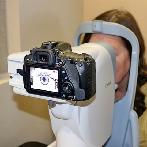
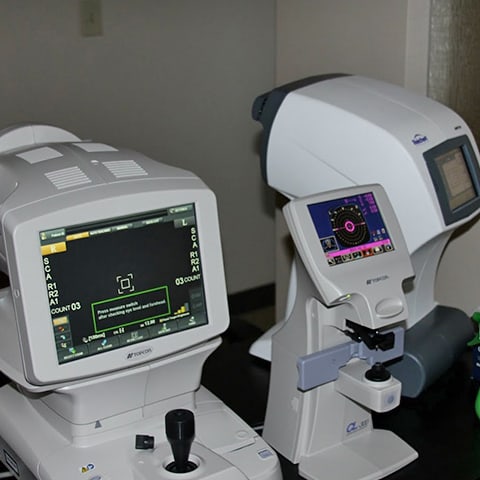
- Eye examinations using advanced technology such as Wavefront digital eye exams
- Pediatric/children eye care (for ages 5 and up)
- Contact lens fitting and lenses including hard-to-fit cases and scleral lenses
Our Location
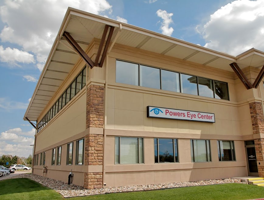
Our Address
Contact Us
- Phone: 719-598-5068
- Email: [email protected]
Clinic Hours
- Monday: 8:00 AM – 5:30 PM
- Tuesday: 8:00 AM – 5:30 PM
- Wednesday: 8:00 AM – 5:30 PM
- Thursday: 8:00 AM – 5:30 PM
- Friday: 8:00 AM – 5:30 PM
- Saturday: Closed
- Sunday: Closed

Our Brands





Our Blogs
Slit Lamp Eye Exam
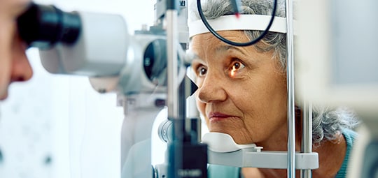
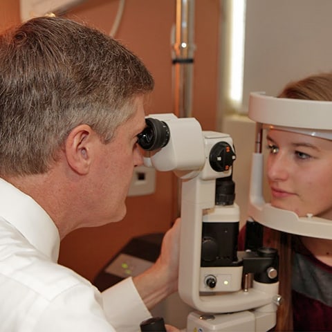
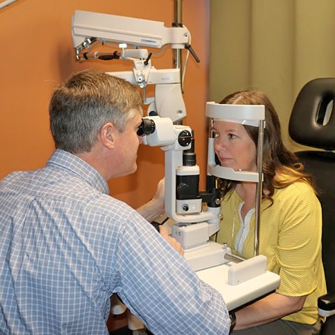
Eye CareWhat is a Slit Lamp Exam? A slit lamp has an opening that allows it to shine a thin “sheet” of light into the eye. The brightness of the light can be adjusted so that the examining doctor either sees the front part of the eye or all the way to the back, where the […]
5 Tips to Protect Your Vision
Eye CareYou use your eyesight every day to work, play and enjoy your life. Too often, people take their vision for granted until the day when something goes wrong. With just a little attention to your vision, you can protect your eyesight so it serves you for the rest of your […]
What are the Types of Glaucoma?
Eye CareThe Glaucoma Research Foundation tells us that more than three million Americans have glaucoma, but only half are aware they have it. Of the people who have glaucoma, at least 90% have open-angle glaucoma, the most common type of glaucoma. However, there are other […]
Slit Lamp Eye Exam
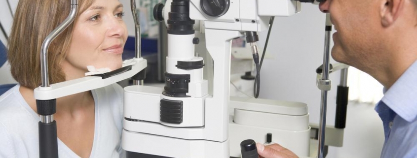
What is a Slit Lamp Exam? A slit lamp has an opening that allows it to shine a thin “sheet” of light into the eye. The brightness of the light can be adjusted so that the examining doctor either sees the front part of the eye or all the way to the back, where the […]
5 Tips to Protect Your Vision
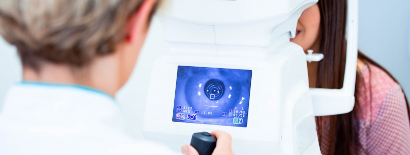
You use your eyesight every day to work, play and enjoy your life. Too often, people take their vision for granted until the day when something goes wrong. With just a little attention to your vision, you can protect your eyesight so it serves you for the rest of your […]
What are the Types of Glaucoma?

The Glaucoma Research Foundation tells us that more than three million Americans have glaucoma, but only half are aware they have it. Of the people who have glaucoma, at least 90% have open-angle glaucoma, the most common type of glaucoma. However, there are other […]


